- Blog /
- September 29, 2021
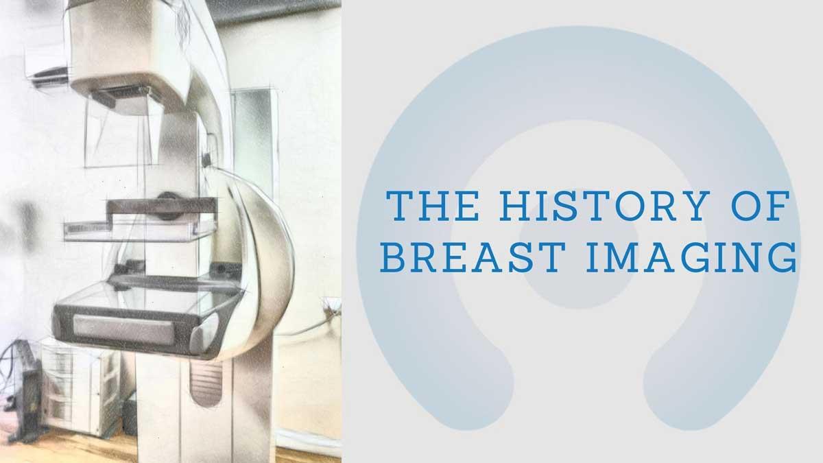
History of Breast Imaging
Breast imaging, as a practice, has gone through numerous technological advances. From direct exposure mammography, xeromammography, and screen-film mammography to the advanced era of field digital mammography and true 3D breast imaging like the Koning Breast CT – the diagnostic technique has come a long way. In addition to these technologies, organized screening and breast imaging reporting and systems continue to shape the development of breast imaging.
Here, we discuss how breast imaging techniques have developed across different eras to help you understand its evolution and importance.
Mammography Screening- The Advent of Breast Imaging
Radiologists previously performed mammography using X-ray tubes in the 1960s. Those exams didn’t have any compression. They used direct-exposure films to capture the imaging in a similar way to chest X-rays. The images were usually very low-shaded, and the tissues close to the wall of the chest appeared “white” because of underexposure.
The next decade saw further advancements in mammography with the invention of screen-film mammography. This made capturing images easier and faster with lower radiation doses. The technology made it easier to spot and see through breast tissue.
More impressments and advancements in screen-film technology related to dedicated mammography took place in the 80s and 90s. This made images exceptionally better.
Over the years, the use of advanced technology for mammography screening became more common. This is because of two factors:
- The result of multiple randomized trials demonstrating the effectiveness of mammography to decrease breast cancer mortality
- The development of useful pre-operative image-guided wires localization technique to obtain tissues diagnosis for lesions detected at screening
Regulating Mammography
In the 90s, further regulations on mammography led to the procedure becoming more common. As breast cancer became a health threat for the public and imaging assessments turned into a basic test, concerns related to variation in the quality of mammography grew.
These concerns resulted in Quality Standards Act for mammography in 1992 to impose uniform standards nationwide. The act required radiologists to provide quality screening and interpretation that can communicate the findings clearly to ensure effective patient care.
New Age of Mammographic Breast Imaging
As the new century began, breast imaging transitioned further with the invention of digital mammography. While advanced digital mammography is the same technique from the perspective of the patient, the new machine makes use of signals to capture images that doctors can read on the computer instead of x-ray films.
Many studies found that when it came to screening dense breasts, digital mammography is better than previous forms of mammography, but it is still difficult to find lesions at their earliest stage. Koning Health realized this and created a true 3D breast imaging option – with no compression. Koning provides better imaging compared to film and digital mammography, and it generates 360-degree 3D images in only ten seconds. The KBCT offers a better view of breast tissue for any type of breast – whether small, large, dense, or fatty.
Summing Up
All in all, breast imaging has come a long way when it comes to developing new technologies, and Koning continues to work to bring the breast imaging industry into the future with its revolutionary technology.
Links
https://www.sir-florida.com/breast-imaging
https://www.koninghealth.com/en/product-solutions/koning-breast-ct
